
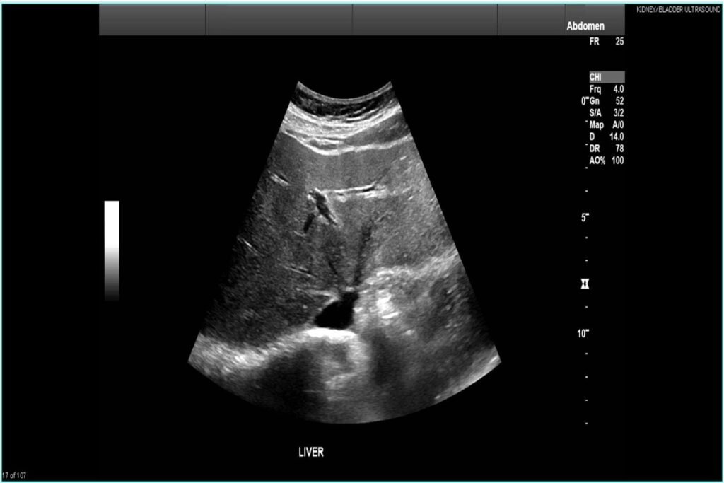
A liver ultrasound is one of the most effective, safe, and non-invasive diagnostic tools available today for evaluating the health of your upper abdomen. Your liver works tirelessly behind the scenes to filter toxins, aid digestion, and store energy, making proactive monitoring essential to your long-term health.
In this comprehensive guide, we will discuss how a liver ultrasound works, what the scan reveals, how it detects fatty liver disease early, what to expect during the procedure, and why booking an appointment with Beverly Hills Ultrasound Center is your best choice for clear, accurate imaging.
What Is a Liver Ultrasound and How Does It Work?
An abdominal imaging examination using a liver ultrasound relies on high-frequency sound waves to capture real-time, detailed visual images of your liver and surrounding tissue. Unlike X-rays or CT scans, ultrasound technology emits zero ionizing radiation, making it exceptionally safe for routine health screenings and repeated monitoring.
During the scan, a trained sonographer applies a water-based gel to your upper right abdomen. A small hand-held device called a transducer is then guided across the skin. The transducer sends sound waves deep into the tissue; as those waves bounce off organ structures, blood vessels, and fluid collections, they travel back to a computer screen to form a sharp, grayscale image.
By analyzing these acoustic reflections, physicians can examine the overall size, shape, outline, and internal tissue texture (echogenicity) of your liver in a matter of minutes.
What Does a Liver Ultrasound Scan Show?
When a specialist evaluates your liver ultrasound output, they receive a detailed visual roadmap of your upper digestive and metabolic systems.
Here is a breakdown of what a sonographer and radiologist look for during your scan:
- Liver Size and Contour: An enlarged liver (hepatomegaly) can indicate inflammation, congestion, or excess fat buildup. Smooth outer borders generally suggest a healthy organ, whereas irregular or nodular surfaces can signal progressive scarring.
- Tissue Texture and Density: Healthy liver tissue exhibits a uniform, mid-level gray tone. Changes in this brightness pattern help physicians distinguish between healthy liver cells and compromised areas.
- Gallbladder and Bile Ducts: Because the liver produces bile to digest fats, the scan examines the gallbladder and bile ducts for gallstones, blockages, or wall thickening.
- Vascular Flow: Advanced ultrasound techniques, including Doppler ultrasound, allow specialists to measure blood circulation through the hepatic arteries and portal vein, ensuring nutrient-dense blood moves smoothly to and from the organ.
- Focal Lesions: The scan readily identifies distinct growths, distinguishing fluid-filled cysts (which are usually benign) from solid masses, hemangiomas, or tumors.
Spotting Fatty Liver Disease Early
Detecting metabolic dysfunction early is where a liver ultrasound truly shines. Metabolic Dysfunction-Associated Steatotic Liver Disease (MASLD), formerly known as Non-Alcoholic Fatty Liver Disease (NAFLD), affects millions of adults worldwide. Fatty liver occurs when excessive fat accumulates within hepatic cells, often without causing any early warning signs or noticeable symptoms.
Key Fact: Fatty liver disease is frequently called a “silent condition” because it can progress for years without causing abdominal pain, fatigue, or abnormal blood work.
When fat builds up in liver tissue, it reflects sound waves differently than normal tissue. On an ultrasound monitor, a healthy liver appears slightly darker than or equal in brightness to the adjacent kidney. However, a liver infiltrated with fat looks noticeably brighter, a visual phenomenon medical professionals call “increased echogenicity” or a “bright liver.”
Grades of Fatty Liver Detected on Ultrasound
| Severity Grade | Ultrasound Visual Characteristics | Clinical Significance |
| Grade 1 (Mild) | Slight increase in liver tissue brightness; blood vessels and diaphragm remain clearly visible. | Highly reversible with targeted lifestyle changes and dietary adjustments. |
| Grade 2 (Moderate) | Moderate brightness; fine details of internal blood vessels become harder to see. | Requires prompt metabolic assessment to prevent tissue inflammation. |
| Grade 3 (Severe) | Marked, intense brightness; deep liver structures and the diaphragm are difficult to visualize. | High risk of advancing to liver inflammation (steatohepatitis) or scarring. |
Detecting fat accumulation at Grade 1 or Grade 2 gives you a crucial window of opportunity. Because the liver possesses remarkable regenerative abilities, early diagnosis via ultrasound allows you and your healthcare team to reverse fat buildup through exercise, nutritional adjustments, and medical guidance before permanent damage occurs.
Other Conditions a Liver Ultrasound Can Identify
While catching fatty liver early is a primary reason patients seek imaging, a comprehensive liver ultrasound is equally vital for uncovering several other abdominal health conditions.
- Liver Cirrhosis and Fibrosis: Chronic inflammation or long-term fatty liver can cause scar tissue to replace healthy cells. Ultrasound scans show this progression through a coarsened, bumpy tissue texture and irregular organ margins.
- Liver Cysts: Simple, fluid-filled sacs are common and usually harmless. An ultrasound easily distinguishes these benign fluid pockets from solid tissue masses.
- Hemangiomas: These non-cancerous collections of blood vessels show up as bright, well-defined spots during an imaging session.
- Jaundice and Bile Duct Obstruction: If you experience yellowing skin or eyes, an ultrasound can instantly pinpoint whether a gallstone or duct blockage is backing up bile fluid.
- Portal Hypertension: By evaluating blood vessel diameter and flow direction using Doppler technology, the scan can check for high blood pressure within the liver’s primary vein system.
How to Prepare for Your Liver Ultrasound Procedure
Getting ready for your liver ultrasound is straightforward, but taking a few simple steps ensures your sonographer gets the clearest possible visual images.
- Fasting Before the Scan: You will typically be asked to fast (no food or drinks other than light sips of water) for 6 to 8 hours prior to your appointment. Fasting reduces gas in your stomach and intestines, which can block sound waves, and causes your gallbladder to fill, making it much easier to evaluate.
- Clothing Choices: Wear a comfortable, two-piece outfit. This allows easy access to your upper abdomen without having to change out of your clothing entirely.
- Medications: You can usually take your standard daily medications with a small sip of water unless your care team specifically advises otherwise.
During the 15- to 30-minute procedure, you will lie comfortably on an examination table. The sonographer will apply warm gel and gently sweep the probe across your ribs and upper stomach. You may be asked to hold your breath for a few seconds at a time; this pushes the liver down past your ribcage for an unobstructed view. The process is completely painless, quiet, and relaxing.
Why Choose Beverly Hills Ultrasound Center for Your Scan?
When it comes to diagnostic imaging, precision, comfort, and professional expertise make all the difference, which is why booking your liver ultrasound at Beverly Hills Ultrasound Center ensures an exceptional healthcare experience.
Located in the heart of Beverly Hills, our state-of-the-art facility features advanced, high-definition ultrasound technology operated by experienced, certified sonographers. We understand that medical appointments can sometimes bring anxiety; that is why we prioritize a calm, luxurious environment where your comfort comes first.
At Beverly Hills Ultrasound Center, you benefit from:
- Cutting-Edge Diagnostic Equipment: Our high-resolution imaging technology captures subtle tissue variations, allowing for earlier detection of fatty liver and small lesions.
- Experienced Clinical Specialists: Our team works alongside board-certified radiologists to deliver rapid, meticulous reports directly to you and your physician.
- Personalized, Patient-Centered Care: We take the time to answer your questions, explain the imaging process, and ensure you feel relaxed throughout your visit.
- Convenient Scheduling: Flexible appointment times fit seamlessly into your busy lifestyle without long wait times.
Taking control of your metabolic and digestive health begins with clear information. Don’t wait for silent symptoms to develop; prioritize your wellness today by scheduling your screening with the trusted experts at Beverly Hills Ultrasound Center.